
Top Medical Research Papers on Neutrophil Extracellular Traps (NETs)

If you're a medical researcher unsure of which direction to take, exploring current hot topics in medical research is a good choice. This blog primarily discusses neutrophil extracellular traps (NETs), a growing research focus. Between 2020 and 2023, the number of projects related to NETs doubled, with funding increasing more than fourfold, reaching a total of nearly $25 million. The number of publications indexed in PubMed has also been steadily rising, with over 400 papers already submitted this year.
So, what are NETs?
NETs are web-like structures secreted by activated neutrophils (a process known as NETosis) and consist of DNA, histones, and antimicrobial proteins. Their formation is one of the active responses to combat infectious damage or non-sterile inflammation. NETs are an essential extracellular defense mechanism of innate immunity.
Next, let's take a closer look at some of the recent research findings. Not only will you gain insight into the role of NETs in different diseases, but you will also learn about different technical approaches and research methods.
Molecular Signature of Neutrophil Extracellular Trap Mediating Disease Module in Idiopathic Inflammatory Myopathy
Journal: Journal of Autoimmunity
Impact Factor: 12.8
Publication Date: May 22, 2024
Research Background:
Neutrophils are the first responders to acute inflammation. During chronic inflammation, the continuous recruitment and activation of neutrophils, along with impaired neutrophil clearance, exacerbate inflammation and cause collateral tissue damage. NET formation is one of the active strategies to combat infectious damage or non-sterile inflammation.
The research team found that NETs play a central role in neutrophil-mediated immunity and share common characteristics across different subtypes of idiopathic inflammatory myopathy (IIM).
Methodology:
Data Collection and Integration: Systematic collection of muscle tissue transcriptomic data from different IIM patient subtypes. Data from different platforms were integrated and standardized to minimize bias.
Differential Gene Expression Analysis: Differentially expressed genes (DEGs) between IIM patients and healthy controls were identified. Time-dependent trends in DEGs were analyzed using ImpulseDE software.
Functional Enrichment Analysis: DEG-related biological processes and pathways were explored using Enrichr, GSEA, and ssGSEA, with GO term categorization and visualization done using REVIGO.
Cell Subpopulation Analysis: xCell software was used for deconvolution of muscle tissue samples to estimate the proportion of different immune cell subpopulations, and differences between IIM subtypes were analyzed.
Gene Regulatory Network (GRN) Analysis: ARACNe algorithm was used to construct GRNs to identify differentially activated regulators and their expression patterns in immune cell subpopulations.
Interferon Stimulated Genes (ISGs) Analysis: ISG scores were calculated to assess interferon signaling activity in IIM subtypes.
Protein-Protein Interaction (PPI) Network Construction: Using human interactome databases, PPI networks were analyzed to identify interactions between DEGs.
Disease Module Detection: TOPAS algorithm was used to identify disease modules from DEGs, revealing core functional gene networks in IIM. Drug targets and their potential roles in these modules were also analyzed.
Network-based Diffusion Scoring: Quantification of the disturbance effect of drug targets on disease modules using kernel functions and diffusion algorithms.
Muscle Injury and Regeneration Model Analysis: Dynamic changes in cell subpopulations and pathways were analyzed in mouse muscle injury and regeneration models, identifying key molecular events related to IIM pathology.
Experimental Results:
Figure 1
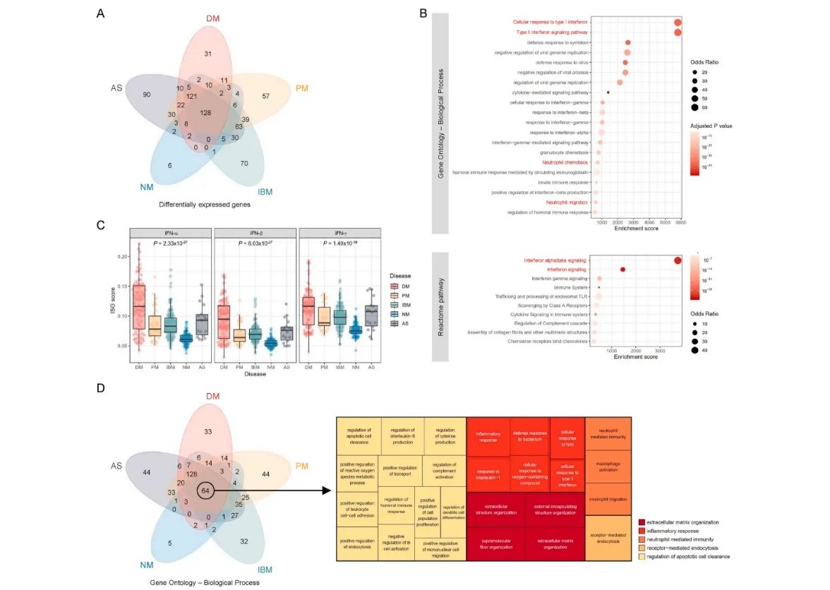
DEGs and Functional Enrichment: Panel A shows a Venn diagram of DEGs from five IIM subgroups, with 128 DEGs identified.
Functional Enrichment: In panel B, enrichment of IFN signaling and neutrophil-mediated immunity pathways in dermatomyositis (DM) was noted.
ISG Scores: Panel C shows significant differences in ISG scores across IIM subtypes, with the lowest score in NM samples.
Shared GO Terms: Panel D identifies 64 shared GO terms across IIM subtypes, categorized into five major clusters: extracellular matrix tissue, inflammatory response, neutrophil-mediated immunity, receptor-mediated endocytosis, and apoptosis regulation.
Characterization of a Predictive Signature for Tumor Microenvironment and Immunotherapy Response in Hepatocellular Carcinoma Involving Neutrophil Extracellular Traps
Journal: Heliyon
Impact Factor: 4.0
Publication Date: May 8, 2024
Research Background:
Hepatocellular carcinoma (HCC) has an increasing incidence and mortality rate. Due to frequent recurrence and metastasis, HCC prognosis remains poor, with a five-year survival rate of less than 18%. The rise of genetic testing and molecular targeted therapies has made gene expression-based prognostic evaluation a hot research topic.
This study aims to predict the prognosis and immunotherapy response of HCC patients using molecular clustering and prognostic signatures of NETs.
Methodology:
Data Analysis: Data was collected from the TCGA and ICGC databases, with the TCGA dataset randomly split into training and testing sets. Data cleaning and preprocessing were done using R.
NETs-related Gene Clustering: Consensus clustering was used to divide HCC patients into two NETs-related subtypes, with survival and clinical features compared. CIBERSORT algorithm was used to analyze immune cell infiltration differences between subtypes.
NETs-related Prognostic Signature Construction: Univariate Cox regression was used to identify prognostic-related DEGs. LASSO regression and multivariate Cox regression were applied to construct a six-gene NETs-related prognostic signature. The prognostic ability was validated using a nomogram model.
Immune Microenvironment Analysis: ESTIMATE algorithm assessed the relative abundance of immune and stromal cells in different risk groups. ssGSEA analyzed immune cell infiltration and immune function differences across risk groups, as well as response differences to immunotherapy.
Gene Function Analysis: Differential gene expression between high- and low-risk groups was analyzed. Gene ontology (GO) and KEGG enrichment analyses identified biological functions and pathways. GSVA was used to analyze potential biological functions.
Drug Sensitivity Analysis: pRRophetics algorithm analyzed differences in drug sensitivity between high- and low-risk groups.
Experimental Validation: qRT-PCR and immunohistochemistry were used to validate NETs-related gene expression in clinical samples. siRNA interference technology was applied to validate the role of HAVCR1 gene in regulating HCC cell proliferation and NET levels.
Research Results:
Figure 5
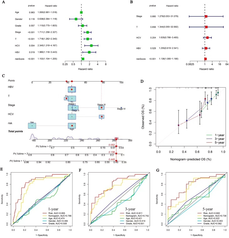
Univariate and Multivariate Analysis: NETs-related features were confirmed as independent prognostic factors (P<0.001).
Nomogram Model: Panel C shows a novel nomogram integrating NETs features and clinical characteristics to enhance prognostic prediction.
Calibration Curve: The calibration curve (panel D) demonstrates strong prognostic predictive value.
ROC Analysis: Panel E-G compares the prognostic prediction ability of the nomogram with clinical features (gender, grading, age) using ROC analysis at 1, 3, and 5 years, showing AUC values of 0.766, 0.742, and 0.739, respectively.
Risk Loci Involved in Giant Cell Arteritis Susceptibility: A Genome-wide Association Study
Journal: The Lancet Rheumatology
Impact Factor: 25.4
Publication Date: May 8, 2024
Research Background:
Previous studies have shown that genome-wide association studies (GWAS) are highly effective in identifying genetic factors contributing to complex diseases such as giant cell arteritis. Searching PubMed with keywords like "giant cell arteritis," "temporal arteritis," and "GWAS" from the database's inception to August 31, 2023, only found one GWAS report detailing three risk loci associated with this vasculitis. These findings were incorporated into this study.
This research represents the largest GWAS and meta-analysis of giant cell arteritis to date, involving 3,498 patients and 15,550 controls, and discovered three new gene loci associated with giant cell arteritis risk, one of which is related to neutrophil extracellular traps (NETs).
Methodology:
Data Collection and Quality Control: Genomic data from giant cell arteritis patients and healthy controls were collected from ten European and North American cohorts. Low-quality single nucleotide polymorphisms (SNPs) and samples were excluded. Data imputation was performed using the TOPMed Imputation server to improve data quality.
Principal Component Analysis: Used to evaluate population stratification and exclude samples with abnormal ancestry.
Association Analysis: Single-variable analysis was performed for each cohort, and results were combined using a fixed-effect inverse variance weighted meta-analysis method.
Functional Analysis:
Cell Enrichment Analysis: Evaluated the enrichment of associated SNPs in various cell types.
Fine Mapping: Bayesian fine mapping was used to identify the most likely causal variants at each risk locus.
Causal Gene Prioritization: Open Targets Genetics tools were used to identify genes related to giant cell arteritis risk.
Drug Repurposing Analysis: Drugs targeting potential causal genes or their interaction proteins were identified using the DrugBank database.
Polygenic Risk Score: A polygenic risk score model was developed to assess the predictive capacity of genetic variants for giant cell arteritis.
Experimental Results:
Figure 1
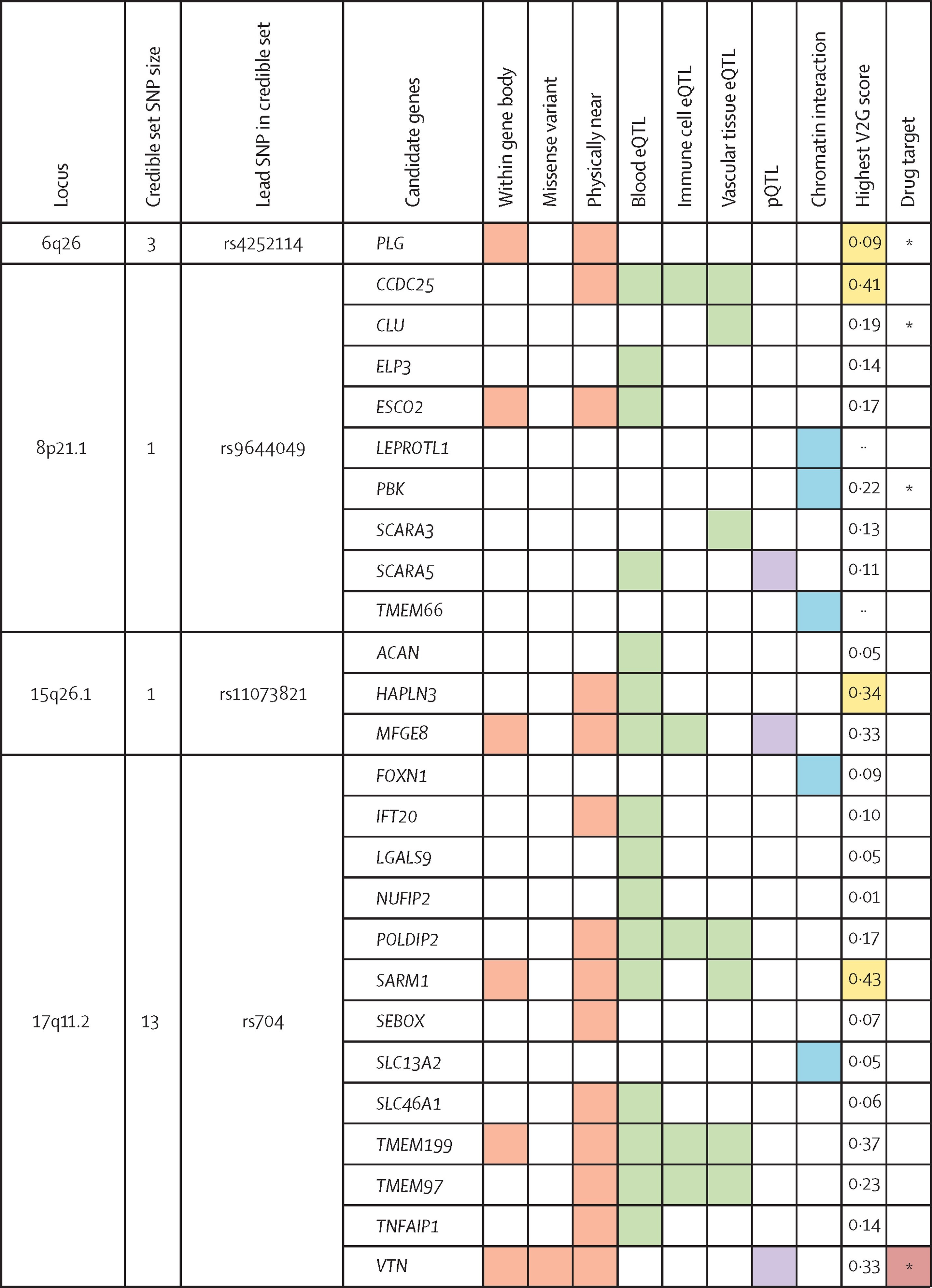
The color in the figure indicates the overlap between SNPs or genes associated with giant cell arteritis and functional annotations. Different colors represent different annotation categories.
Using FUMA gene mapping and Open Targets Genetics, 26 candidate genes were identified, with PLG (chromosome 6), CCDC25 (chromosome 8), MFGE8 (chromosome 15), and VTN (chromosome 17) being the most supported.
The study also found an intronic variant of CCDC25, with the risk allele associated with increased CCDC25 expression in immune cells. CCDC25 encodes a transmembrane receptor involved in NETs formation, triggering ILK-β-parvin pathway activation and enhancing cell motility. This pathway, which is associated with cancer metastasis, may play a role in enhancing immune cell recruitment in pathological arterial tissues in giant cell arteritis. Notably, NETs-induced MMP-9 expression by monocytes has been observed in granulomatosis with polyangiitis, another form of vasculitis affecting small blood vessels.
Accelerate Your Research with Scifocus: AI-Powered Support for Efficient Data Analysis
In medical research, especially in cutting-edge fields like NETs, quickly and accurately accessing, analyzing, and synthesizing vast amounts of scientific data is crucial. With Scifocus, researchers can leverage AI-driven tools for literature search, data analysis, and research design, accelerating the pace of their work. The intelligent recommendations and research assistance offered by Scifocus help you easily find the latest studies and advancements in your field, providing deeper insights into the molecular mechanisms of NETs and their applications across different diseases. Whether you're a novice or an experienced researcher, Scifocus offers powerful support to enhance your research efficiency.
References
Moon, S.-J., Jung, S. M., Baek, I.-W., Park, K.-S., & Kim, K.-J. (2024). Molecular signature of neutrophil extracellular trap mediating disease module in idiopathic inflammatory myopathy. Journal of Autoimmunity. https://doi.org/10.1016/j.jaut.2023.103063
Z Yuan, X Yang, Z Hu, Y Gao, P Yan, F Zheng, Y Guo, & X Wang, J Zhou. (2024). Characterization of a predictive signature for tumor microenvironment and immunotherapy response in hepatocellular carcinoma involving neutrophil extracellular traps. Heliyon, 10(1), Article e06858. https://doi.org/10.1016/j.heliyon.2024.e33955
Borrego-Yaniz, G., Ortiz-Fernández, L., Madrid-Paredes, A., Kerick, M., Hernández-Rodríguez, J., Mackie, S. L., et al. (2024). Risk loci involved in giant cell arteritis susceptibility: A genome-wide association study. The Lancet Rheumatology, 6(2), 123-134. https://doi.org/10.1016/S2665-9913(24)00064-X
Did you like this article? Explore a few more related posts.
Start Your Research Journey With Scifocus Today
Create your free Scifocus account today and take your research to the next level. Experience the difference firsthand—your journey to academic excellence starts here.