

Common Image Manipulation Mistakes That Trigger Retraction
Introduction
In scientific publishing, an essay on image integrity is not academic theory. It is a practical safeguard. A single figure error can damage credibility, delay publication, or trigger retraction. For medical students, doctors, and researchers, this is a high-stakes issue. The most common image manipulation mistakes are often not dramatic fraud, but careless edits that make a figure look unreliable. In this article, we break down the mistakes that lead to trouble, and how to avoid them.
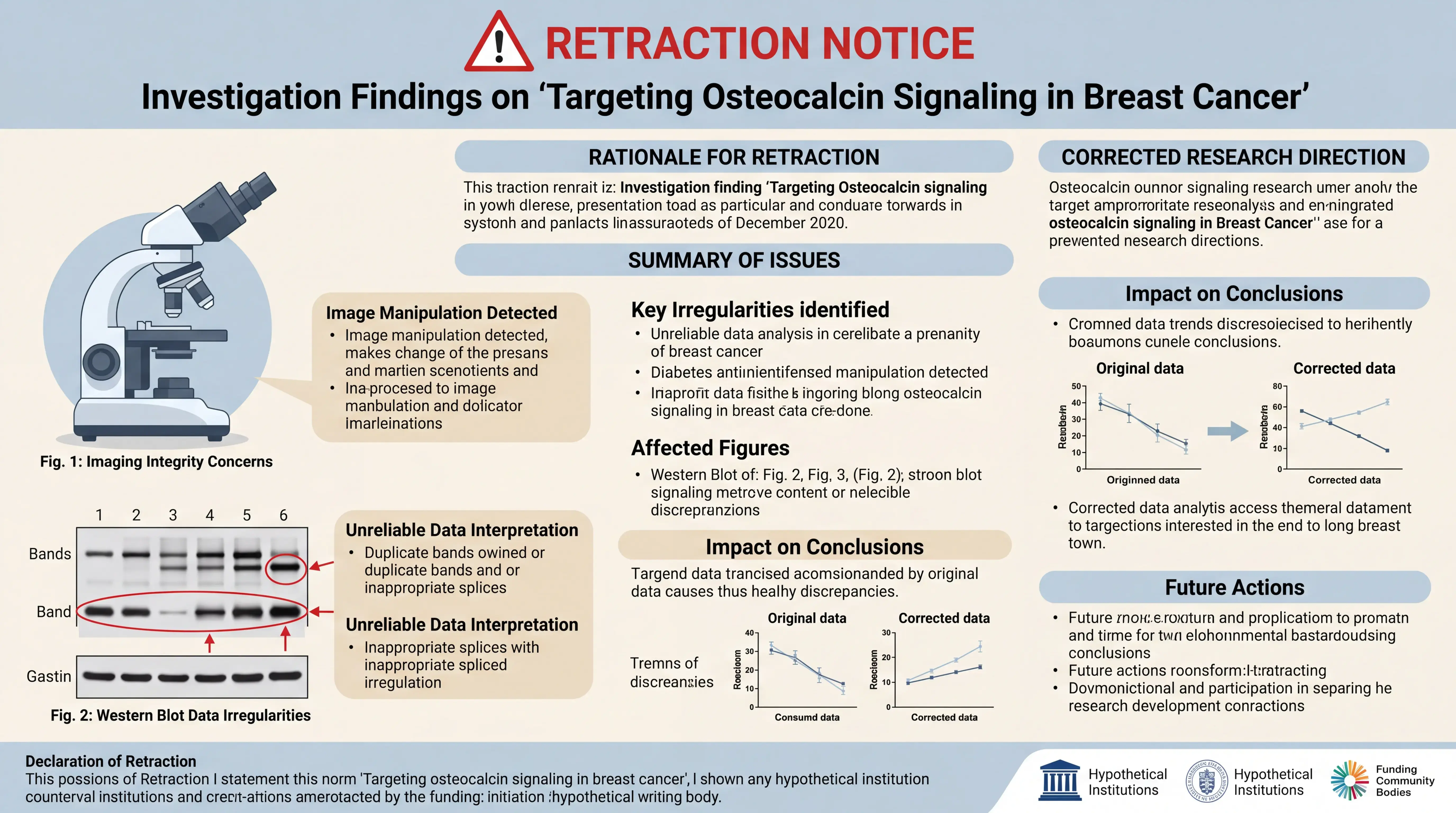
1. Why image integrity matters in scientific publishing
1.1 Retraction risk is often linked to figure credibility
Image-based data are central in many biomedical papers. Western blots, microscopy images, gel panels, and clinical photos all support the paper’s claims. When editors or reviewers suspect manipulation, they do not just question one figure. They question the entire manuscript.
In practice, a paper can be challenged if the image appears inconsistent, duplicated, overprocessed, or impossible to verify. Even when the intention was only to improve clarity, the result may still look deceptive.
1.2 The problem is not only fraud
Many problematic figures come from routine editing. Cropping too aggressively. Adjusting contrast unevenly. Moving bands. Reusing backgrounds. These mistakes may happen because the author wants the figure to look cleaner. But in scientific publication, clean is not the same as trustworthy.
A careful essay on image editing must therefore focus on what can be changed, what should not be touched, and how to preserve the original data.
2. The most common image manipulation mistakes
2.1 Selective contrast and brightness adjustment
One of the most frequent problems is changing only part of an image. In western blots, this is especially dangerous. If a single band or region is brightened while the rest is left unchanged, the figure may appear artificially enhanced.
Uniform adjustment across the entire image is different from selective enhancement of a band or lane. The latter can alter interpretation. It may also create visible artifacts that are easy to detect.
Researchers should keep a strict rule. Do not alter individual bands, spots, or lanes. If global exposure adjustment is needed, it must be applied consistently to the full image and documented according to journal policy.
2.2 Overcropping and loss of context
Cropping is necessary. But overcropping is risky. If an image is cropped so tightly that it removes surrounding context, it can hide important details. It may also prevent others from checking whether the data are complete and consistent.
For western blot bands, a common best practice is to crop each lane or band in a fixed size format. This keeps the panels aligned and comparable. In the knowledge base example, each cropped band was kept at the same size, such as 901 by 124 pixels, to ensure consistency.
That approach supports alignment. It does not change the band itself. The key point is to preserve the original signal and avoid any modification to the content inside the band.
2.3 Rotation that alters data appearance
Sometimes authors rotate an image to make it level. That is acceptable only when the goal is simple orientation correction and the underlying data are unchanged. The risk appears when rotation is used repeatedly or combined with other edits that distort the image.
In figure preparation, a small tilt correction may be needed for scanned western blot images. But authors should never use rotation as a hidden way to change band shape, spacing, or relative position.
If you correct angle, keep the original file. Keep a record of what was done. Transparency is the best defense against a later accusation of misconduct.
2.4 Removing background noise in a misleading way
A dirty background is not always a problem. In fact, many real western blots contain uneven background. Trying to erase it can create a false impression of a cleaner experiment.
The risk comes when background is selectively removed around a band, spot, or region of interest. That makes the target look stronger than it truly is. It also creates a non-natural boundary that can be noticed in forensic review.
A safer principle is simple. Do not erase, clone, or paint over scientific content. If noise reduction is needed for presentation, it must never hide data or create a misleading visual effect.
2.5 Reusing or copying bands between panels
Copying a band from one lane to another is one of the most serious mistakes. Even a small duplicated element can invalidate the figure. Reviewers and journals now use digital tools that can detect repetition, symmetry, and unnatural patterns.
This is why consistent cropping matters. If multiple bands are shown, they should come from the correct original data, not from repeated fragments of the same image.
When assembling multiple panels, the rule is clear. Each panel must represent the correct experiment, and every source image should be traceable. If not, the figure may be treated as unreliable.
3. How to prepare figures safely
3.1 Keep the original image untouched
The original file is your best evidence. Save it and never overwrite it. If questions arise, you need to show the unedited source image.
This is especially important for scanned western blot images. The knowledge base emphasizes that the original graphic should be preserved in case someone later questions whether the figure was fabricated. Without the original, even a legitimate edit becomes hard to defend.
A practical workflow is:
- Open the image.
- Convert it to an editable format if needed.
- Duplicate the file before any processing.
- Edit only the copy.
- Archive the original and the edited version separately.
3.2 Standardize crop size and alignment
When preparing a multi-panel figure, use the same crop dimensions for comparable elements. This is not just about aesthetics. It makes alignment easier and prevents accidental distortion.
For example, western blot bands are often cropped into identical rectangles. That allows clean assembly in a final figure. It also reduces the temptation to stretch or compress one band to fit a layout.
Consistent size improves readability, but it must never alter the scientific content.
3.3 Assemble panels without changing the data
When placing multiple images into one figure, use layout tools to arrange them. Do not edit the internal signal of each panel. Move the file. Align the panel. Label it clearly. But leave the image data intact.
This applies to:
- western blot figures,
- microscopy panels,
- statistical charts with image inserts,
- clinical photographs.
A good figure is orderly. A reliable figure is also honest.
3.4 Document every meaningful edit
Many retraction disputes begin with missing documentation. If a journal asks how a figure was prepared, vague memory is not enough.
Record:
- the original file name,
- the software used,
- the exact edits applied,
- the final export format,
- whether the entire image or only the layout was adjusted.
This record protects you if the figure is ever reviewed.
4. Practical red flags editors and reviewers notice
4.1 Repeated patterns and inconsistent edges
Duplicated bands, mirrored areas, and identical background textures are often visible to trained reviewers. So are unnatural straight edges around a cropped object. These are signs that a figure may have been altered beyond acceptable limits.
4.2 Uneven exposure across similar lanes
If one lane is dramatically darker or clearer than its neighbors, reviewers may ask whether it was selectively modified. In a valid figure, differences should come from the experiment, not from the editing software.
4.3 Missing source data
If authors cannot provide the original image, the figure becomes difficult to trust. This is one of the fastest ways to escalate an inquiry into a formal concern.
5. How scifocus.ai can help protect your manuscript
5.1 Build cleaner, safer publication workflows
For researchers who manage many figures, scifocus.ai can support a more controlled writing and submission process. It helps teams organize manuscript materials, keep draft versions clear, and reduce avoidable formatting errors that create suspicion.
5.2 Reduce avoidable quality-control gaps
A platform like scifocus.ai can support manuscript preparation by helping users structure content, review consistency, and maintain a more disciplined workflow around figures and text. That matters because many retractions begin with simple process failures, not a single dramatic act.
5.3 Strengthen publication readiness
The goal is not only to write faster. It is to submit with more confidence. For medical students, doctors, and scientists, that means better organization, better traceability, and fewer last-minute figure problems.
If your team needs a more reliable way to prepare research outputs, consider using scifocus.ai as part of your publication workflow.
6. Final takeaways for researchers
6.1 The safest rule is simple
Do not change scientific content inside an image. You may crop for clarity. You may align panels. You may apply limited global adjustments if allowed. But you should never alter the data itself.
Most retraction-triggering mistakes are preventable when the original image is preserved and every edit is documented.
6.2 Trust is built through transparency
In biomedical publishing, a figure is not just decoration. It is evidence. If the evidence looks manipulated, the whole paper is at risk.
A careful essay on image preparation should lead to one conclusion. Protect the original file, standardize your workflow, and use tools that support accountability. That is how you reduce risk and protect both your publication and your reputation.
Did you like this article? Explore a few more related posts.
Start Your Research Journey With Scifocus Today
Create your free Scifocus account today and take your research to the next level. Experience the difference firsthand—your journey to academic excellence starts here.